Home > Exhibits > Teaching Tools > Early Years (1910s-1950s)


Teaching Tools

Early Years (1910s-1950s)

Moulages

The predecessors of plastic anatomical models used today, moulages have been in use since the Renaissance. Physicians worked with artists to develop these life-like simulations of various diseases and conditions. Many materials were used to create moulages, but the most popular and durable was wax due to its ability to cast directly from the human body.
 
The moulages displayed here are molded wax on a plaster base. They were created by David Rhea, a laboratory assistant in charge of tissue processing for the Department of Pathology. Rhea worked under the direction of Perry Tollman, MD, chair of the Department of Pathology (1948-1954) and later dean of the College of Medicine (1954-1964).


Top

Stereocards

A predecessor of holographic displays, stereoscopes enabled the viewer to see an image in 3D. Invented in 1832 by Sir Charles Wheatstone, it included mirrors and prisms to customize viewing. By the 1840s, the stereoscope became accessible to the masses, thanks to the creation of a portable lenticular stereoscope and the advent of commercially successful daguerreotype photography. By the early 1900s, physicians used stereoscopes to provide their students with 3D studies of human anatomy outside the laboratory.

Top

Microscope & Student Study Slides


Microscopes have evolved considerably since their creation in the early 17th century. High tech microscopes on campus today far surpass this 1940s UNMC student microscope.


Slide-making is a focal point of pre-med science courses and pathology studies. UNMC’s cytotechnology program became available to international learners in 2019. This program provides virtual education in microscopy to students all over the world who may not have had access to labs and slides for the study of human cell diagnostics.


Top

Teaching Slides

The “magic lantern” was an early image projector created in the 1600s, initially used for entertainment. In the 1800s, lantern slides became a useful way to project and share information in a classroom. Professors made the slides using images or specimens, a transfer paper that allowed typing directly onto the slide, and two pieces of glass wrapped with tape. Robert Spencer Wigton, MD, made these slides when he was a professor of neurology and psychiatry at UNMC from 1946 to 1971.

Top

Medical Illustrations

Medical illustrations are a significant educational tool as they enhance health sciences instruction, authors' publications, and materials used to educate the public.

August Frederick Jonas, MD

From the McGoogan Health Sciences Library Special Collections and Archives

August F. Jonas, MD, Collection

Early Medical Illustrations

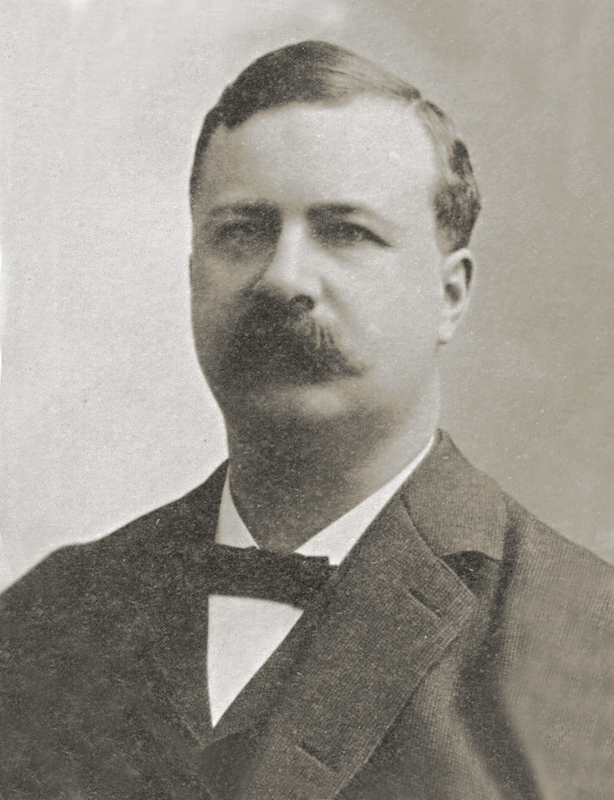
August Frederick Jonas, MD, was born in Wisconsin in 1858. He started studying medicine at the age of 14 and received his degree from Bennett Medical College in Chicago in 1877. Upon graduation, he practiced in Sauk City, Wisconsin, for five years before going abroad in 1882 for additional medical studies at Ludwig Maximillian University in Munich, Bavaria. He graduated in 1884 and conducted post-graduate studies in Vienna, Berlin, and Paris before returning to the United States.


Dr. Jonas arrived in Omaha in 1887, where he organized the surgical department at Methodist Hospital and was named chief surgeon. Dr. Jonas taught surgery at UNMC starting in 1892 and served as chair of the Department of Surgery from 1919 until his retirement in 1930. He served as dean of the college from 1898 to 1902. During World War I, Captain Jonas served as special medical aid to the Governor of Nebraska and was chair of the organizational committee for Base Hospital No. 49 and the Omaha Ambulance Company.


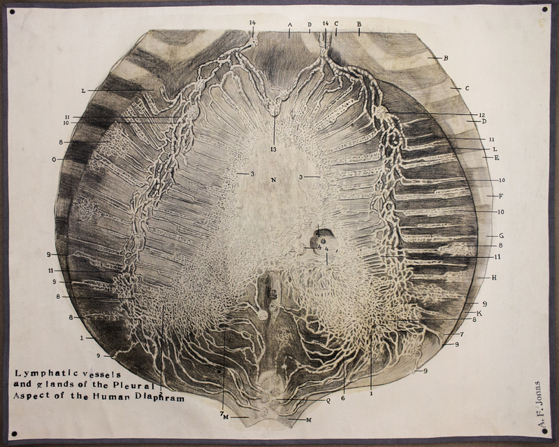
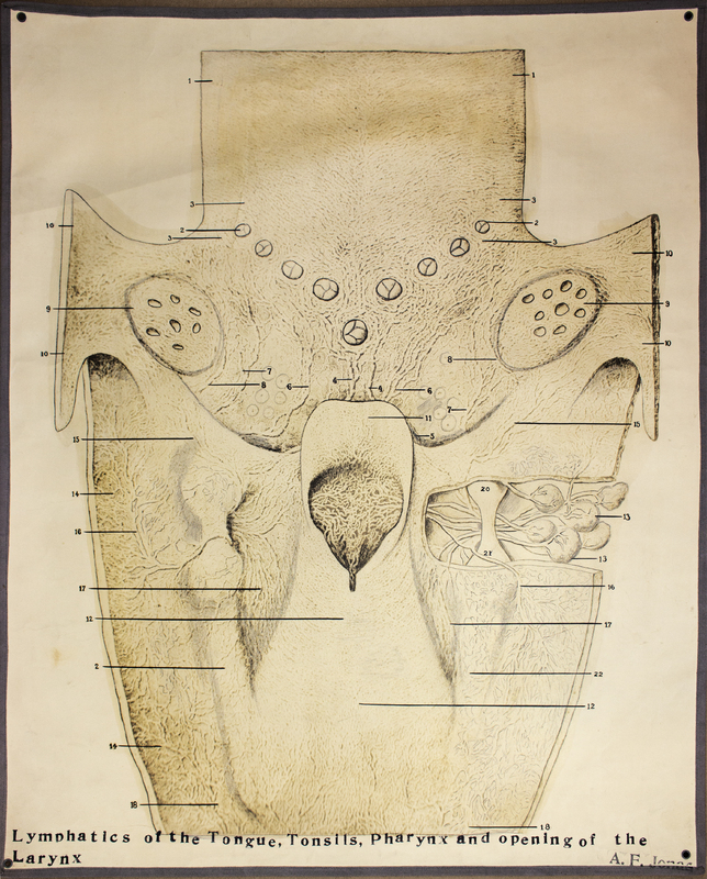
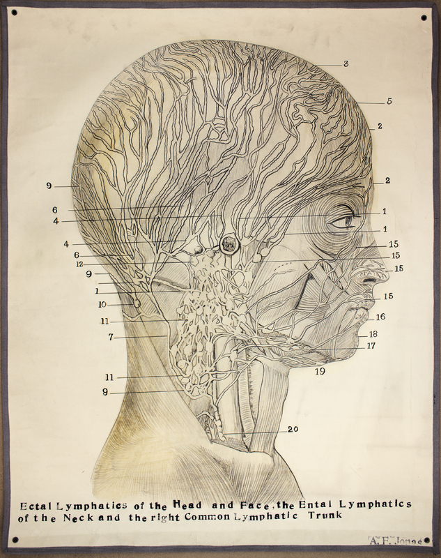
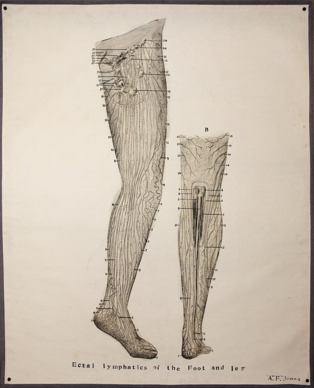
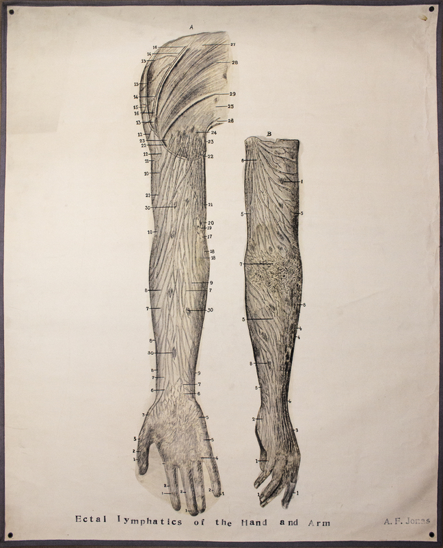
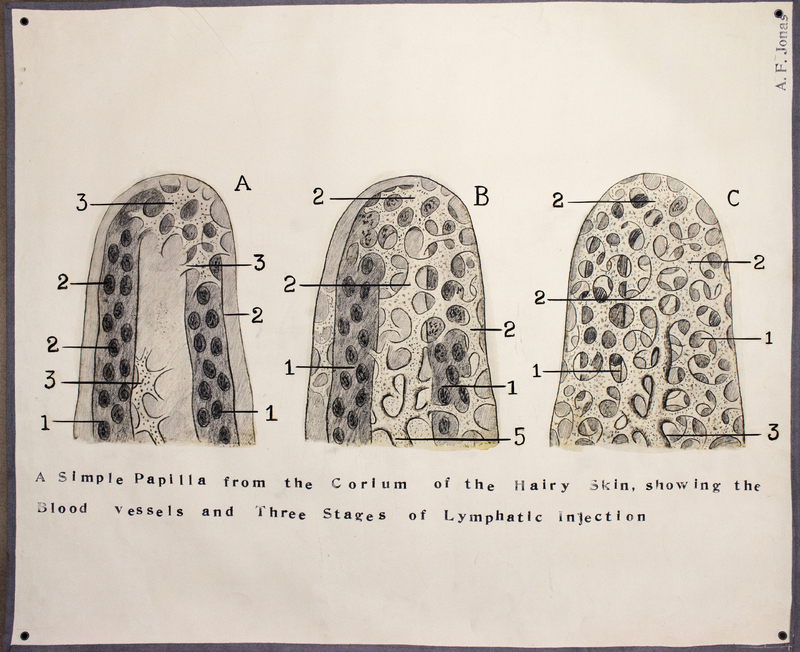
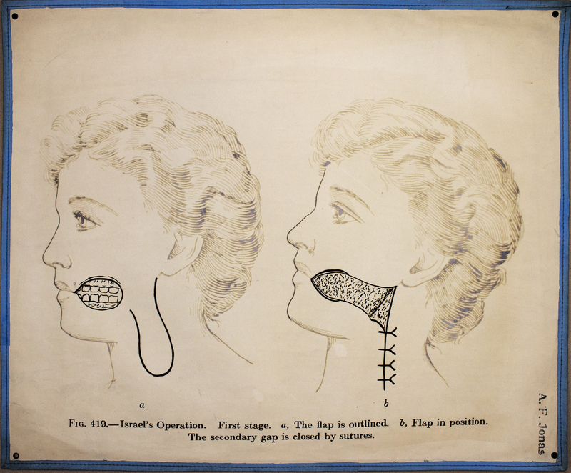
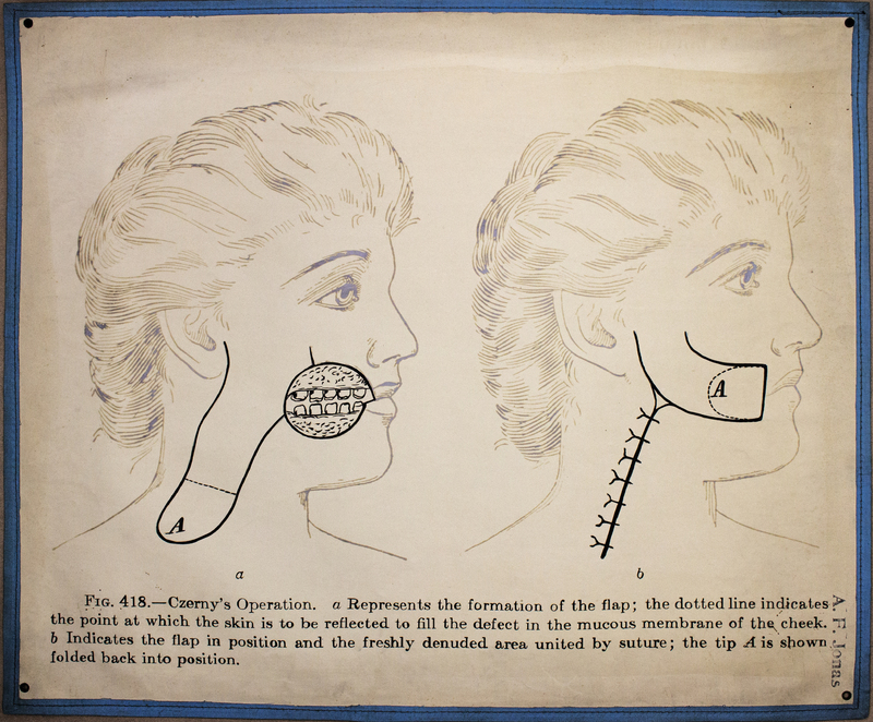
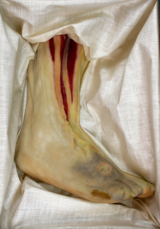
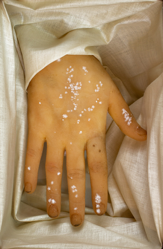
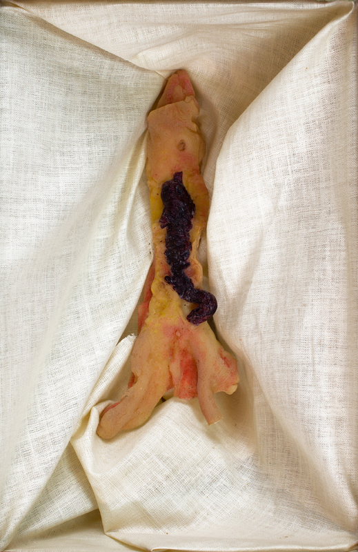
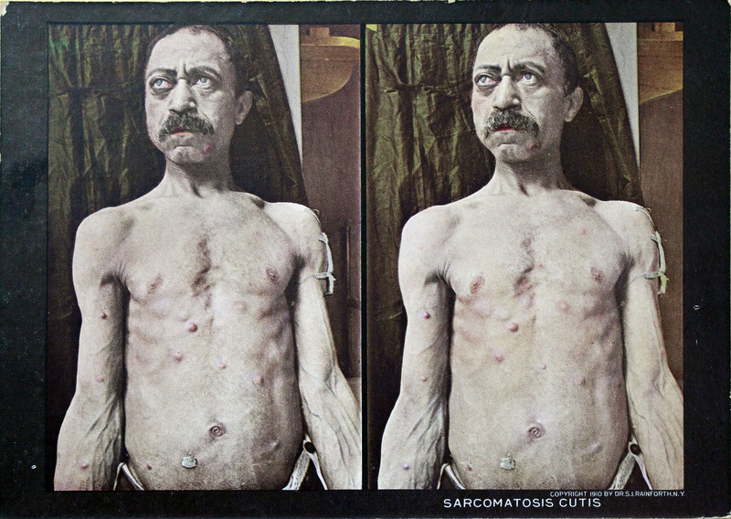
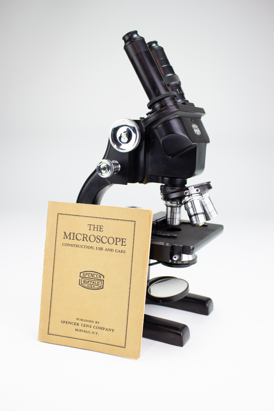
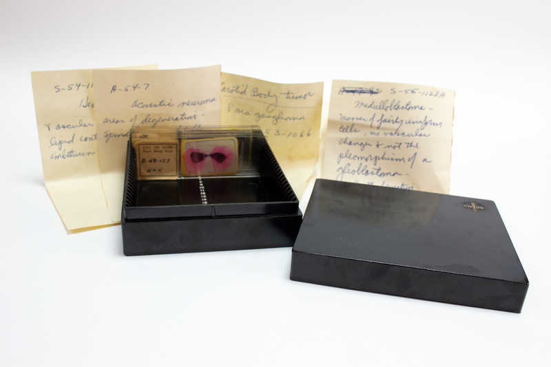
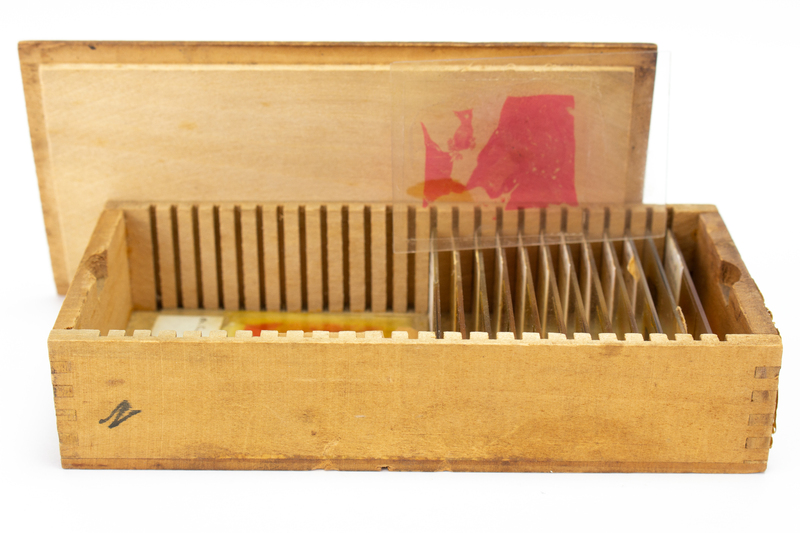
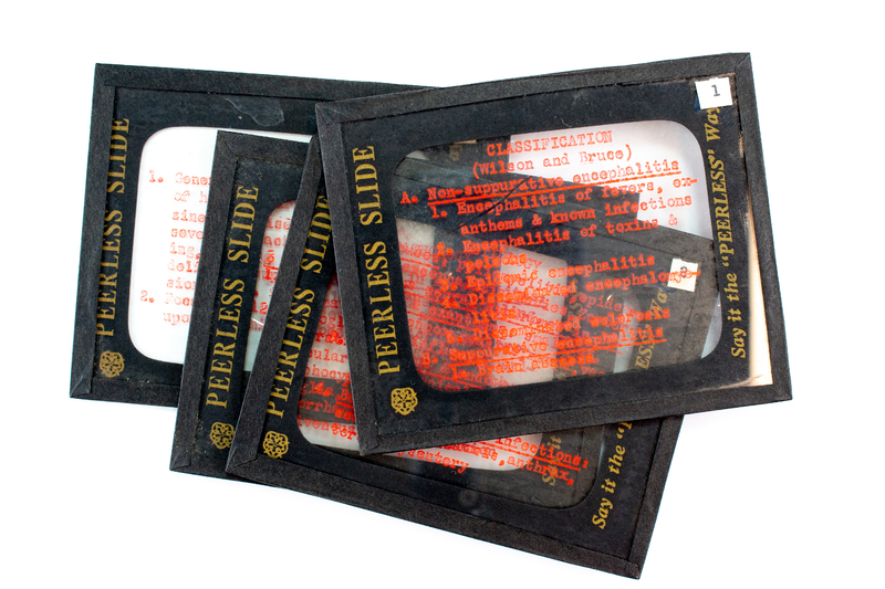
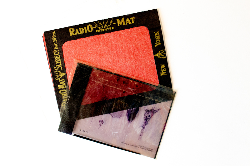
Dr. Jonas used his collection of surgical illustrations in the classroom, providing students with another avenue outside of the anatomy lab to learn about the human body.


Top